Examinations

You can have the following examinations carried out in our practice:

Magnetic resonance tomography (MRI)

learn more

Magnetic resonance imaging (also called magnetic resonance tomography or MRI) uses strong magnetic fields to create sectional images of the inside of the body with the aid of a computer. The state-of-the-art 3 Tesla high-field MRI used in our centre offers the advantage of an open design, i.e. wide opening – short magnet – with the best image quality. In combination with a sophisticated acoustic and visual entertainment system, the examination is in most cases easily possible even for patients with claustrophobia. MRI examinations are not recommended for patients with pacemakers, cochlear implants (implanted hearing aids) and non-removable insulin pumps because of the strong magnetic fields.

Mamma-MRI

learn more

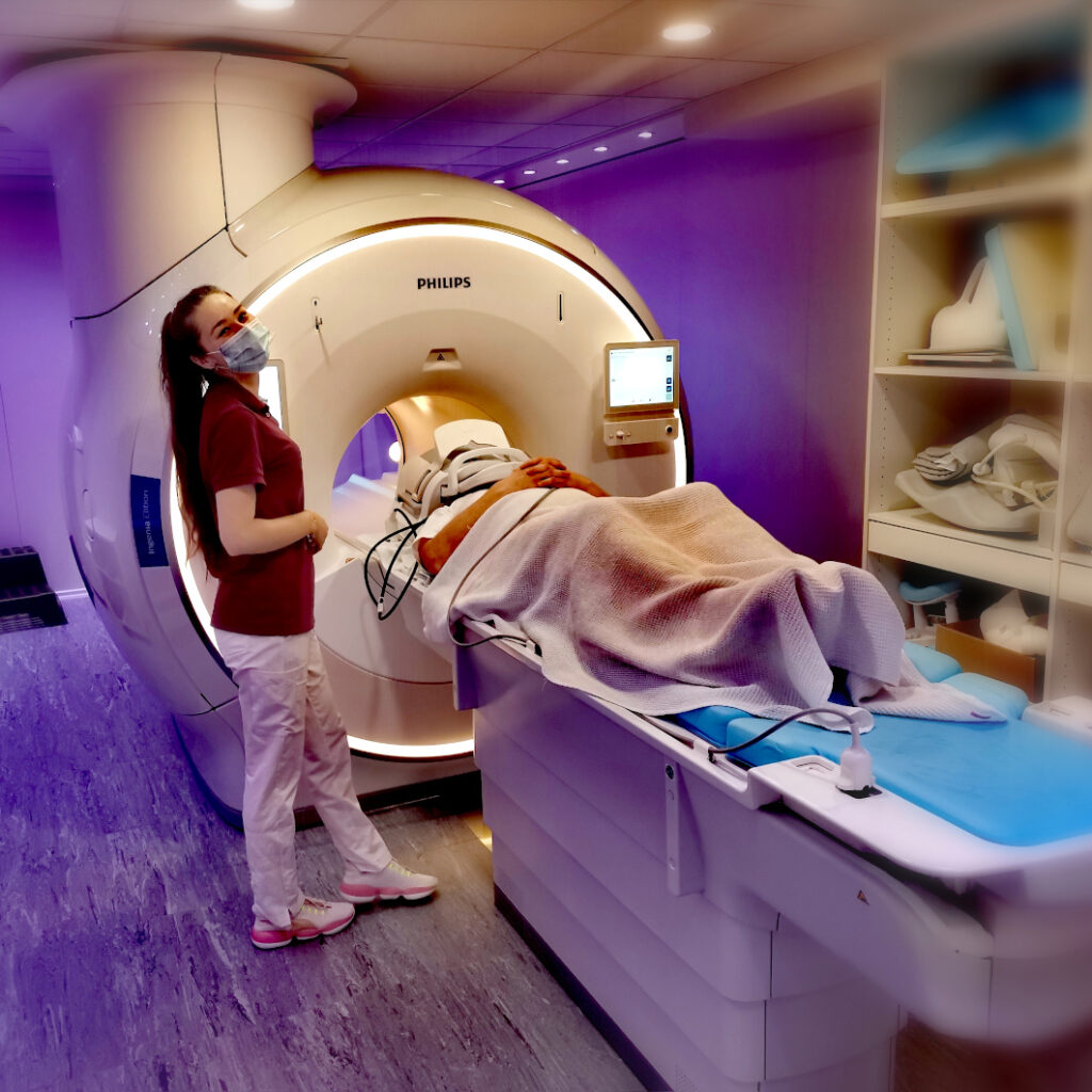
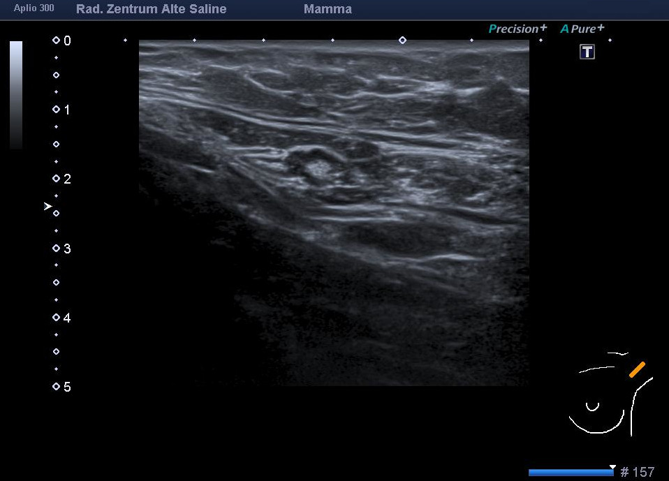
An MRI examination of the breast, the so-called breast MRI, is a very sensitive method with which even small breast carcinomas and tumour precursors (DCIS) can be diagnosed.
This method can be used to detect tumours that are not yet visible in X-ray mammography and ultrasound. The detection rate of breast MRI is about 90 %.

The costs of breast MRI are usually covered by private health insurance companies without any problems.

Up to now, the statutory health insurance companies in Germany have only paid for breast MRI in exceptional cases; unfortunately, we cannot offer you any settlement via your statutory health insurance company.

If you would like to have the examination performed as a “self-payer”, this is of course also possible. Please do not hesitate to contact us, we will be happy to advise you.

Prostate MRI (mpMRI)

learn more

The multiparametric MRI examination of the prostate makes it possible to detect prostate cancer at an early stage or to exclude the presence of prostate cancer with a high degree of certainty. It is currently considered the most sensitive procedure for detecting changes that are suspicious for prostate cancer.

Private health insurances usually cover the costs of an mpMRI without any problems.

Unfortunately, the statutory health insurance funds in Germany do not currently pay for mpMRI, despite the recommendations in the prostate cancer guideline, i.e. it is not a standard benefit. However, patients with statutory health insurance can have the examination carried out by us as “self-payers”. We will be happy to advise you on this.

Computertomography

learn more

Using X-rays, sectional images of the inside of the body are created by a computer. Compared to conventional X-rays, this method allows a much more detailed assessment of the inside of the body. The image information can also be significantly increased by using an X-ray contrast medium and subsequent image processing. The area of examination depends on the question to be clarified. In principle, the method can be used on the entire body. By using a new modern computed tomography system with powerful image computers that make use of artificial intelligence (AI), the examinations can be performed with comparatively low radiation exposure.

Digital Mammography

learn more

Mammography is the imaging of breast tissue using X-rays. Thanks to a new digital mammography system and modern image processing technology, only a relatively low radiation exposure is required for this examination in our practice. In addition, we attach great importance to an examination that is as painless as possible for the patient. The images obtained allow the doctor to recognise structural abnormalities and the smallest calcifications, which are often the only indication of malignant changes in the breast tissue.

Digital X-Ray

learn more

This is an imaging procedure that uses X-rays to show mainly bone structures and also the lungs. This so-called conventional X-ray is increasingly being replaced by cross-sectional imaging (CT and MRI).

Niere

Ultrasound

learn more

In ultrasound, sound waves are emitted into the body from a transmitter guided by the doctor’s hand, reflected differently inside the body and sent back to the transmitter, which also acts as a receiver. A computer “translates” the reflected sound waves into an image on which the structures inside the body can be recognised. In addition to imaging the mammary gland, the ultrasound examination is often used in our practice to examine the abdominal organs and the thyroid gland.

Mammary ultrasound

learn more

Sonography (=ultrasound) is now a firmly established and effective procedure for clarifying benign and malignant changes in breast tissue. In combination with a high-frequency transducer and colour-coded duplex sonography, even very small tumours can be detected with a relatively high degree of certainty, even in dense parts of the glandular tissue. Mammary sonography has been an integral part of the diagnostic spectrum in our practice for many years.